Appendix 8: Anatomic locations of ventricular septal defects
Appendix 8 for the New Zealand Obstetric Ultrasound Guidelines.
The inlet septum separates the two atrioventricular (AV) valves.
The outlet septum includes the conal and infundibular septum and is the region below the arterial valves and above the crista supraventricularis.
The membranous septum is the thin portion in the left ventricular outflow tract (LVOT) just beneath the aortic valve and under the crista supraventricularis.
The muscular septum is the thickest portion of the septum, extending from the tricuspid valve attachments to the apex.
Located in the outflow tract beneath the aortic valve and under the supraventricular crest. May be sub-classified as:
Located under the pulmonary valve and above the supraventricular crest.
Located in the muscular septum, may be mid-muscular, apical or multiple (‘Swiss cheese’ septum).
Located posterior to the septal leaflet of the tricuspid valve. Visualised in four-chamber (4Ch) view near the AV valves.
May be difficult to differentiate from a mild complete or partial atrioventricular septal defect (AVSD).
Drop-out artefact on apex up greyscale imaging and colour Doppler ‘bleeding’ artefact can simulate an inlet ventricular septal defect (VSD).
The diagnosis can be aided by assessing in a lateral or transverse plane.

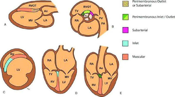

(A) long-axis view, (B) short-axis view at aortic valve level, (C) short-axis view through ventricles, (D) 4Ch view, (E) LVOT view.
Source: Morrison and McMahon 2018.