Marginal cord insertion


  • The umbilical cord inserts into the margin of the placenta, usually defined as within 20 mm of the placental edge (although sometimes defined as <10 mm).
  • Occurs in approximately 7 percent of singleton pregnancies but about 25 percent of twin pregnancies (particularly monochorionic twins).
  • Occasionally a marginal cord insertion may progress into velamentous due to trophotropism later in the pregnancy.
  • It may be associated with complications, such as fetal growth restriction and preterm birth.
  • Follow-up growth assessment may be required.

Image A6.1

Marginal cord insertion Marginal cord insertion

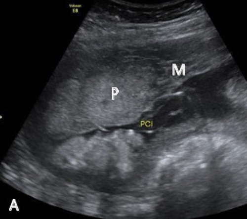
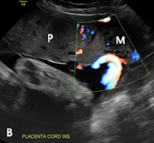
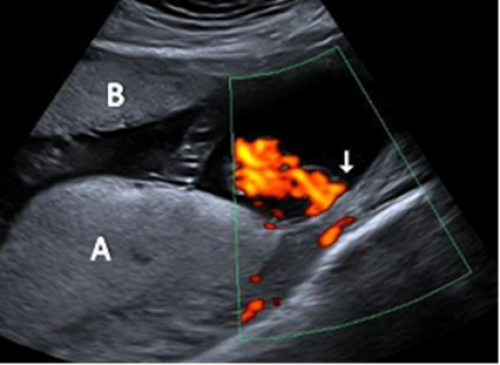
Marginal cord insertion on greyscale (A) and colour Doppler (B) imaging.
Placenta (P), myometrium (M).

Velamentous cord insertion


  • In this condition, the umbilical cord inserts into the fetal (chorioamniotic) membranes, coursing within the membranes to the placenta (between the amnion and the chorion). The exposed vessels are not protected by Wharton’s jelly and are vulnerable to rupture.
  • Velamentous cord insertion has been associated with an increased risk of adverse perinatal outcomes and is associated with vasa previa (where fetal vessels traverse the internal os in front of the leading fetal part); a condition that is associated with high perinatal mortality when not diagnosed prenatally.
  • It may also be associated with placenta previa earlier in pregnancy.
  • Abnormal cord insertion may be associated with fetal growth restriction and intrauterine fetal demise.
  • There is an increased incidence in twin pregnancies, particularly monochorionic twins.
  • Growth surveillance is recommended.

Image 6.2

Velamentous cord insertion Velamentous cord insertion

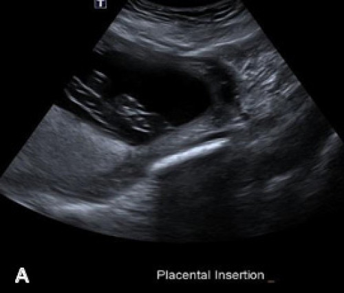
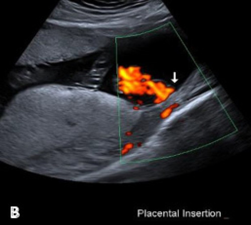
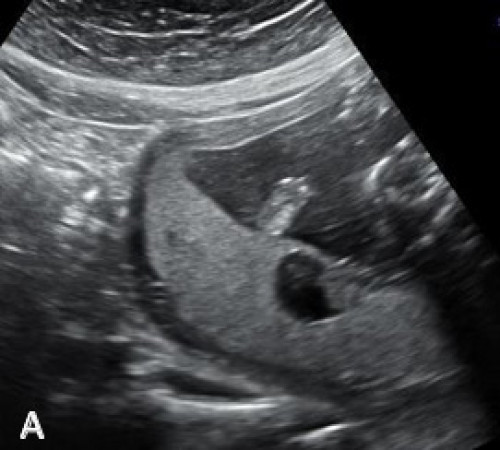
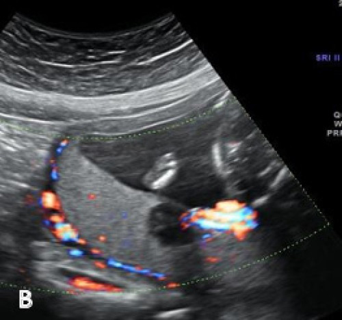
Velamentous cord insertion on greyscale imaging (A) and with colour Doppler (B).

Succenturiate lobe


  • A variant in placental morphology, when there is one or more smaller accessory placental lobe separate from the main disc of the placenta.
  • Occurs in approximately 2 in every 1,000 pregnancies.
  • When a succenturiate lobe is identified, it is important to assess for the location of connecting vessels, and particularly, vasa previa.
  • In a bilobed placenta, the two lobes are of similar size.


Associations/complications


  • Type II vasa previa
  • Increased incidence of postpartum haemorrhage due to retained placental tissue.

Image 6.3

Succenturiate lobe Succenturiate lobe

Main placental lobe posteriorly (A) and smaller anterior succenturiate lobe (B), associated with a velamentous cord insertion.

Chorioangioma


  • Benign tumour within the placenta
  • Occurs in approximately 0.5–1 percent of pregnancies
  • Most commonly diagnosed in the second trimester
  • Usually asymptomatic, unless large
  • On ultrasound, the lesion is a relatively well-defined hypo- or hyperechoic mass distinct from adjacent placenta, typically near the cord insertion. Internal vascularity may be evident on colour Doppler examination
  • Large lesions (>5 cm), may be associated with complications such as polyhydramnios, preterm labour, fetal cardiomegaly, growth restriction, pre‑eclampsia or abruption.

Image A6.4

Chorioangioma Chorioangioma

Heterogeneous hypoechoic lesion within the posterior placenta on greyscale imaging (A) and with colour Doppler (B), showing its location adjacent to the placental cord insertion.