Normal early intrauterine pregnancy
Learn more about Normal early intrauterine pregnancy guidelines
These guidelines were published in 2019 and are awaiting review, due 2022. Some content may be outdated.
Structures generally develop in the following predictable sequence.
The timing of structure development is also fairly predictable.
Correlate with ultrasound appearances (see below) and refer to local clinical guidelines.
Figure 1: βhCG chart
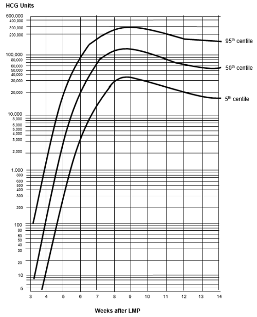
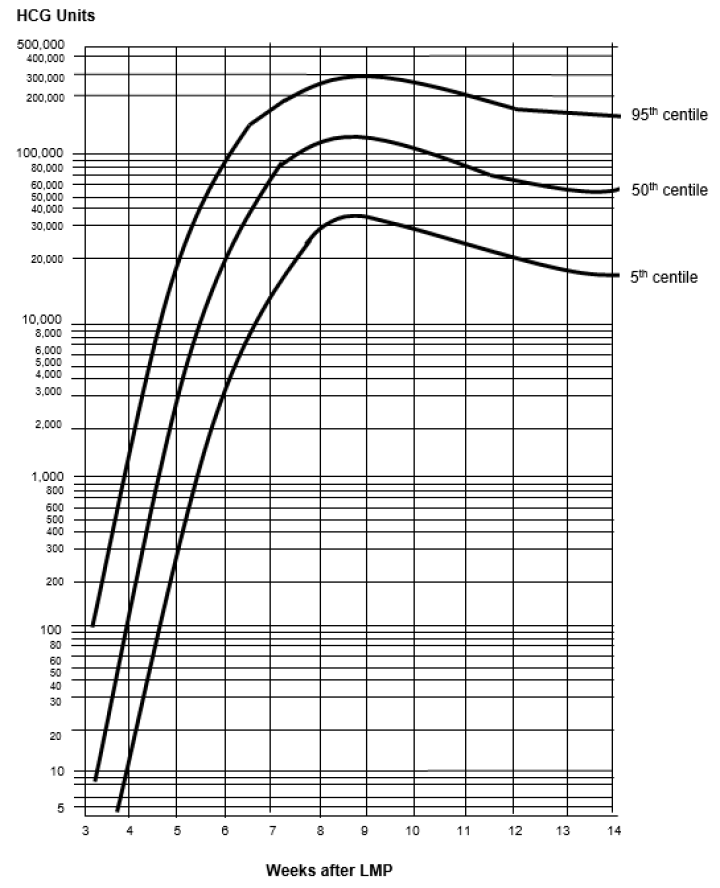

Source: Canterbury SCL, April 2019.
The presence of a yolk sac within the intrauterine gestational sac confirms an intrauterine pregnancy and essentially excludes ectopic pregnancy.
Heterotopic pregnancy is very rare but should be considered if there are suggestive ultrasound features, particularly in the setting of assisted reproductive technology.
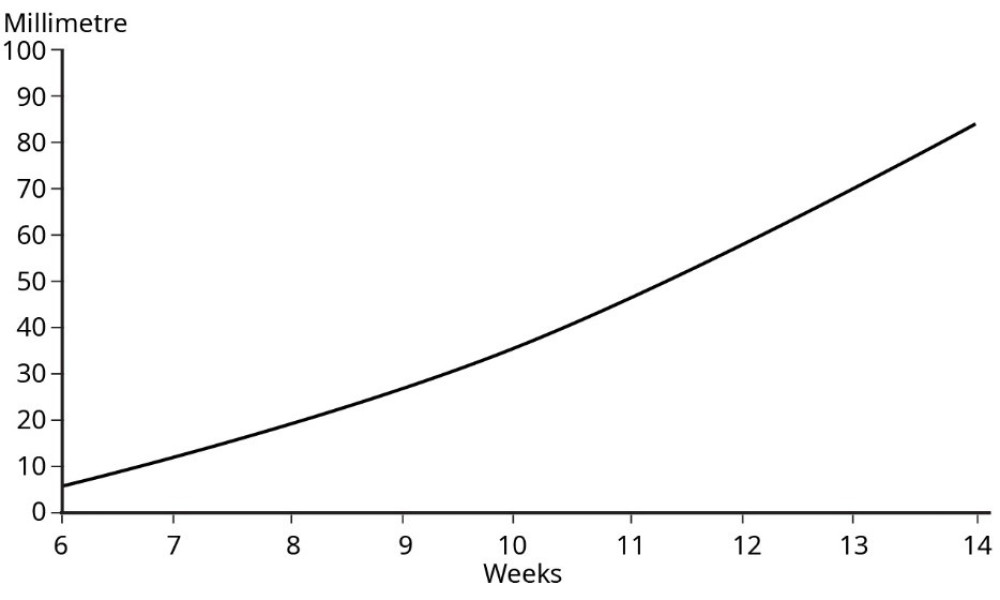
Growth is approximately 1.2 mm per day, but may be less in a normally developing pregnancy. Interval growth of CRL alone should not be used as a determinant of pregnancy loss.


Growth is approximately 1.2 mm per day, but may be less in a normally developing pregnancy. Interval growth of CRL alone should not be used as a determinant of pregnancy loss.
Data source: Westerway et al 2000.
Embryonic cardiac activity should always be visualised with a CRL ≥7 mm.
Slow embryonic heart rate of <80 bpm may suggest a guarded prognosis for the pregnancy.
Suggest a follow-up scan if clinically appropriate.