These guidelines were published in 2019 and are awaiting review, due 2022. Some content may be outdated.

Doppler assessment should follow the national guidelines recommended in the SGA (PDF, 2.9 MB) and Obstetric Doppler guidelines (PDF, 3.7 MB) (NZMFMN 2014a and 2014b respectively).


Routine fetal Doppler is not indicated in a normal, low-risk pregnancy.


For more detail, see Doppler in Obstetrics (PDF, 16.2 MB) (Nicolaides et al 2002).

Indications for Doppler


  1. Suspected fetal growth restriction / compromise / hypoxia
    • Umbilical artery (UA PI), with the addition of middle cerebral artery (MCA PI), ductus venosus (DV PI) as per the SGA and Obstetric Doppler guidelines (NZMFMN 2014a and 2014b respectively).
  2. Screening for severe early onset FGR or pre-eclampsia in high-risk women
    • Note: Routine screening in low-risk women is not currently recommended in New Zealand.
    • Uterine artery Doppler at 20–24 weeks.
  3. Assessing for fetal anaemia
    • MCA PSV or V-max.
  4. MCDA twins (covered in the section under the next chapter Twin pregnancy)

1. Suspected fetal growth restriction / compromise / hypoxia


Umbilical artery pulsatility index (UA PI)


Indications


  • Suspected or known SGA fetus
  • EFW on customised chart (eg, GROW) is <10th percentile
  • EFW on customised chart is dropping percentiles by ≥30 percent
  • AC on the population scan chart is <5th percentile
  • Discrepancy (≥30 percent) between the HC and AC percentile with lower AC percentile
  • Maternal hypertensive disorders, for example, pre-eclampsia
  • Decreased fetal movements.


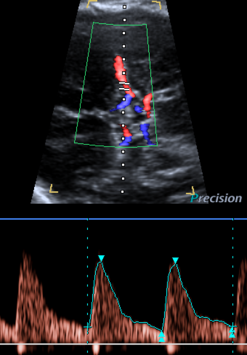
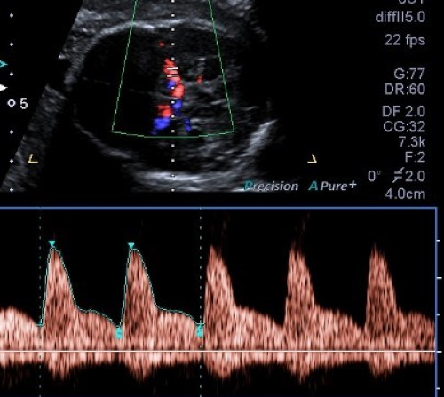
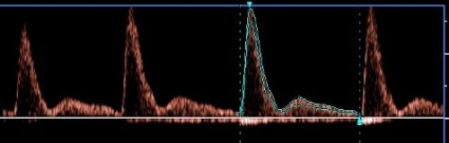
The abnormal UA waveform shows progressive high-resistance flow with reduced, absent and then reversed diastolic flow.


>95th percentile is abnormal.


Middle cerebral artery pulsatility index (MCA PI)


Indications


  • SGA/FGR with abnormal UA PI at any gestation
  • SGA/FGR with normal UA PI after 34 weeks gestation.


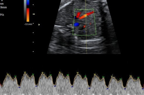
Abnormal MCA PI indicates fetal compensation by redistribution of fetal blood flow to the brain (‘brain sparing’) and shows progressive low-resistance flow with increasing diastolic flow.


<5th percentile is abnormal.


Cerebroplacental ratio (CPR)


If MCA PI assessment is performed, the CPR should be calculated and recorded.

The CPR is the ratio of MCA PI and UA PI (ie, MCA PI divided by UA PI).

<5th percentile is abnormal.


Ductus venosus pulsatility index (DV PI)


DV can be technically challenging to measure and should only be performed by experienced individuals.


Indications to perform DV PI (if possible)


An abnormal DV PI (± absent or reversed atrial (A) wave) indicates fetal cardiac decompensation.


>95th percentile is abnormal.

2. Screening for severe early onset FGR or pre‑eclampsia in high-risk women


Mean uterine artery pulsatility index


Indications


  • Screening of women at high risk of early pre-eclampsia or early SGA at 20 or 24 weeks
  • If abnormal at 20 weeks, repeat at 24 weeks
  • Early onset FGR
  • Current hypertensive disorder in pregnancy
  • Full assessment of suspected SGA or PET in pregnancy.


Abnormal mean uterine artery pulsatility index

  • >95th percentile is abnormal.
  • Notching after 24 weeks is abnormal.

3. Assessing for fetal anaemia


Middle cerebral artery peak systolic velocity (MCA PSV)


Indications


  • Maternal-fetal alloimmunisation
  • Any suspicion of fetal anaemia
  • Unexplained hydrops
  • MCDA twins >24 weeks gestation
  • MCDA twins with known or suspected TTTS or twin anaemia-polycythaemia sequence (TAPS).


>1.5 MoM is abnormal.

Doppler technique


See New Zealand Obstetric Doppler Guideline (PDF, 16.2 MB) (NZMFMN 2014b).


Umbilical artery pulsatility index


  • If the PI is within normal range, only sample one of the umbilical arteries.
  • If the PI is abnormal, sample both umbilical arteries and use the more normal (lower) value.


Middle cerebral artery pulsatility index


  • Perform assessment during fetal quiescence.
  • Start with the BPD view and move caudally to visualise the butterfly shape of suprasellar cisterns and the sphenoid.
  • Assess the MCA that is closer to the transducer.
  • Move anteriorly and angle back to align the MCA flow direction with the Doppler beam.


Ductus venosus pulsatility index


  • Perform assessment during fetal quiescence.
  • Sagittal and transverse approaches are acceptable as long as the Doppler angle is 0–60 degrees.
  • Use colour Doppler to identify the DV at the end of the umbilical vein.
  • If PI >95th percentile, assess umbilical vein for pulsatility.


Middle cerebral artery peak systolic velocity


  • As above for MCA PI but with an ideal interrogation angle of 0 degrees.
  • An interrogation angle of 30 degrees or less is acceptable but requires angle correction.
  • If PSV >1.5 MoM, obtain three high-quality samples and use the highest value.

Doppler waveforms: Normal and abnormal


Umbilical artery Doppler

Image 33

Umbilical artery Doppler Umbilical artery Doppler

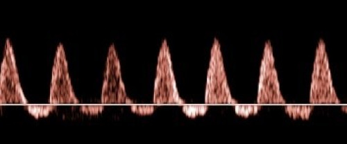
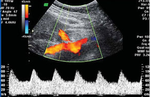
Normal umbilical artery Doppler: Low resistance.

Image 34

Umbilical artery Doppler Umbilical artery Doppler

Abnormal: High PI / resistive index (high resistance trace).

Image 35

Umbilical artery Doppler Umbilical artery Doppler

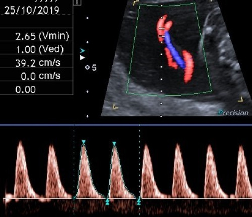
Abnormal: Very high PI / resistive index with absent end-diastolic flow and pulsatility in the umbilical vein.

Image 36

Umbilical artery Doppler Umbilical artery Doppler

Abnormal: Reversed end-diastolic flow.

Image 37

Middle cerebral artery Doppler pulsatility index Middle cerebral artery Doppler pulsatility index

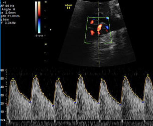
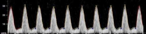
Normal MCA Doppler: High resistance with low diastolic flow velocities.

Image 38

Middle cerebral artery Doppler pulsatility index Middle cerebral artery Doppler pulsatility index

Abnormal: Low-resistance MCA waveform with high diastolic velocities.

Ductus venosus Doppler

Image 39

Ductus venosus Doppler Ductus venosus Doppler

Normal waveform with A wave above baseline.

Image 40

Ductus venosus Doppler Ductus venosus Doppler

Abnormal waveform: A wave below baseline and increased pulsatility.

Image 41

Uterine artery Doppler Uterine artery Doppler

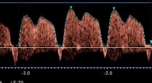
Normal low impedance / low resistance waveform with high diastolic flow and no notch.

Image 42

Uterine artery Doppler Uterine artery Doppler

Normal diastolic flow (normal impedance to flow) but early diastolic notching.

Image 43

Uterine artery Doppler Uterine artery Doppler

Abnormal waveform: Increased PI, with reduced diastolic flow and early diastolic notching.

Middle cerebral artery peak systolic velocity (MCA PSV)

Image 44

Doppler scan showing middle cerebral artery peak systolic velocity Doppler scan showing middle cerebral artery peak systolic velocity

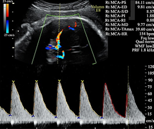
Elevated MCA PSV, well above 1.5 MoM.

Reporting guide and recommendations


  • Report the PI value and whether it is normal or abnormal for each Doppler performed.
  • Report the MoM for MCA PSV, if performed.

Reporting alerts

Urgent


  • Critically abnormal Doppler, eg, umbilical artery absent or reversed end-diastolic flow
  • MCA PSV >1.5 MoM

Same day


  • Mildly abnormal Doppler, requiring follow-up.